Optomap
|
optomap Retinal Examinations ("optomap"), provide eye-care practitioners with clinically useful information that facilitates the early detection of disorders and diseases evidenced in the retina.
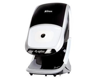
Optos' core product is the retinal image produced by the Panoramic200 Scanning Laser Ophthalmoscope device: the optomap retinal image.
The optomap retinal image gives eye-care professionals a much larger view (200 degrees) of the back of the eye your retina than conventional eye exam equipment. The images can be taken without dilating your pupils a very common procedure which is uncomfortable and inconvenient for many people.
|
The optomap image is captured in less than a second and is immediately available for doctor and patient to review. The optomap Retinal Exam offers many clinical, practice and patient benefits.
The optomap image is displayed immediately after being taken, allowing the eye care professional to review it quickly and if necessary, refer you to a retinal specialist. Using the internet, the image can be sent anywhere in the world for a specialist to review.
Each optomap image is as individual as fingerprints or DNA and can provide eye care professionals with a unique view of your health very quickly and comfortably. The optomap image is captured in less than one second and is immediately available for you and your doctor to review.
The optomap retinal image offers many advantages including:
- An ultra-widefield view of the retina
- Comfortable and quick image capture
- Non-invasive
- Helps you understand your eye health
- Provides permanent records for future comparison
- The optomap technology does not require pupil dilation, however the decision to dilate or not is a medical decision to be made by your health care professional
- Patient can resume normal activities immediately.
For more information visit the company's web site: www.optos.com